Question:
Which one of the following options correctly matches the ECG signals in Panel-1 with the cardiac conditions in Panel-2?

Which one of the following options correctly matches the ECG signals in Panel-1 with the cardiac conditions in Panel-2?
Show Hint
Normal = clean regular P-QRS; AV block = delayed/dropped conduction; atrial flutter = fast sawtooth waves.
Updated On: Aug 7, 2026
- X-(i); Y-(iii); Z-(ii)
- X-(i); Y-(ii); Z-(iii)
- X-(ii); Y-(i); Z-(iii)
- X-(ii); Y-(iii); Z-(i)
Show Solution

Verified By Collegedunia
The Correct Option is A
Solution and Explanation
Step 1: Understanding the Question.
The figure shows three ECG traces, X, Y and Z, and asks us to match each one to a cardiac condition: normal condition, atrial flutter, or atrioventricular (AV) block.
Step 2: Key Concept.
Each condition leaves a distinct fingerprint on the ECG. A normal ECG shows one clean P wave before every QRS complex, at a steady rate. In AV block, the electrical signal from the atria is delayed or fails to reach the ventricles on some beats, so the trace shows disturbed timing between atrial and ventricular activity, such as a stretched interval or a dropped QRS, while the overall rhythm still looks broadly regular otherwise. In atrial flutter, the atria fire abnormally fast, near 250 to 350 times a minute, producing a rapid, repetitive sawtooth pattern of flutter waves riding under the QRS complexes.
Step 3: Detailed Explanation.
Trace X shows evenly spaced beats with a normal-looking P-QRS-T pattern and no extra waves or missed beats, so X is the normal condition, item (i).
Trace Y shows disturbed timing between the atrial and ventricular activity, consistent with delayed or blocked conduction from atria to ventricles, the signature of atrioventricular block, item (iii).
Trace Z shows the fast, jagged, repeating sawtooth-like oscillations typical of the rapid atrial firing seen in atrial flutter, item (ii).
Step 4: Final Answer.
X matches (i) normal condition, Y matches (iii) atrioventricular block, and Z matches (ii) atrial flutter. This is option (A).
\[ \boxed{\text{X-(i); Y-(iii); Z-(ii)}} \]
The figure shows three ECG traces, X, Y and Z, and asks us to match each one to a cardiac condition: normal condition, atrial flutter, or atrioventricular (AV) block.
Step 2: Key Concept.
Each condition leaves a distinct fingerprint on the ECG. A normal ECG shows one clean P wave before every QRS complex, at a steady rate. In AV block, the electrical signal from the atria is delayed or fails to reach the ventricles on some beats, so the trace shows disturbed timing between atrial and ventricular activity, such as a stretched interval or a dropped QRS, while the overall rhythm still looks broadly regular otherwise. In atrial flutter, the atria fire abnormally fast, near 250 to 350 times a minute, producing a rapid, repetitive sawtooth pattern of flutter waves riding under the QRS complexes.
Step 3: Detailed Explanation.
Trace X shows evenly spaced beats with a normal-looking P-QRS-T pattern and no extra waves or missed beats, so X is the normal condition, item (i).
Trace Y shows disturbed timing between the atrial and ventricular activity, consistent with delayed or blocked conduction from atria to ventricles, the signature of atrioventricular block, item (iii).
Trace Z shows the fast, jagged, repeating sawtooth-like oscillations typical of the rapid atrial firing seen in atrial flutter, item (ii).
Step 4: Final Answer.
X matches (i) normal condition, Y matches (iii) atrioventricular block, and Z matches (ii) atrial flutter. This is option (A).
\[ \boxed{\text{X-(i); Y-(iii); Z-(ii)}} \]
Was this answer helpful?
0
0
Top GATE BM Human Anatomy and Physiology Questions
- During the repolarization phase of a neuron, the cell is brought back to the resting potential by the action of a Sodium-Potassium pump. Which one of the following statements is TRUE for the active transport of Na+ and K+ ions through the cell membrane ?
- The cardiac rhythm in a healthy human heart originates from ______.
- Consider the following layers: subcutaneous fat, viable epidermis, stratum corneum, and dermis. Which one of the following represents the correct sequence of the layers from skin surface to within ?
- Macrophages that are resident in the liver are ________.
- Match the different chambers/locations of a healthy human heart in Column-1 to the ranges of diastolic pressures in Column-2.
Column-1 Column-2 (P) Arterial (I) 2-6 mm Hg (Q) Pulmonary artery (II) 8-12 mm Hg (R) Right ventricle (III) 60-80 mm Hg
View More Questions
Top GATE BM Cardiovascular system Questions
- The cardiac rhythm in a healthy human heart originates from ______.
- Match the different chambers/locations of a healthy human heart in Column-1 to the ranges of diastolic pressures in Column-2.
Column-1 Column-2 (P) Arterial (I) 2-6 mm Hg (Q) Pulmonary artery (II) 8-12 mm Hg (R) Right ventricle (III) 60-80 mm Hg - The end-diastolic ventricular volume is found to be 125 mL and the end-systolic ventricular volume is found to be 50 mL. If the heart rate is 65 beats/minute, what is the cardiac output in liters per minute? (Rounded off to 2 decimal places.)
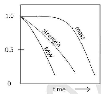
- An orthopaedic implant when monitored over 6 months showed the following normalized curves for polymer molecular weight (MW), mass of implant and mechanical strength. Among the choices, what is the most probable reason for the observed changes ?
- In an attempt to integrate engineered tissue with native tissue, three samples of engineered tissue, X, Y, Z, with identical material properties, were co-cultured adjacent to three different native tissues (bone, cartilage and liver). The adhesive strengths of X, Y, Z were observed after 8 weeks as follows.
Adhesive strength for X = 150 kPa, Y= 250 kPa, Z= 350 kPa
Match the native tissue that were used to co-culture X, Y and Z from the following.
I : Liver Tissue
II : Articular Cartilage
III: Devitalized Bone
View More Questions
Top GATE BM Questions
- What is the value of the following complex line integral counter-clockwise ?
\(\oint_{|z|=3}\frac{8}{z(z-2)(z-4)}dz\) - To solve the equation x = 2 cos x using Newton-Raphson's method, which one of the following iterations should be used ?
- During the repolarization phase of a neuron, the cell is brought back to the resting potential by the action of a Sodium-Potassium pump. Which one of the following statements is TRUE for the active transport of Na+ and K+ ions through the cell membrane ?
- The cardiac rhythm in a healthy human heart originates from ______.
- Which one of the following events is NOT typically encountered in diagnostic X-ray projection radiography ?
View More Questions