Question:

Which thickened nerve is shown here?
Which thickened nerve is shown here?
Show Hint
A superficial nerve crossing the sternocleidomastoid toward the ear and parotid is the greater auricular nerve.
Updated On: Jun 23, 2026
- Facial Nerve
- Greater auricular nerve
- Vagus Nerve
- Glossopharyngeal Nerve
Show Solution
Verified By Collegedunia
The Correct Option is B
Solution and Explanation
Step 1: The thickened nerve shown crossing the sternocleidomastoid in the neck is the greater auricular nerve, a cutaneous branch of the cervical plexus.
Step 2: Its anatomy identifies it. The greater auricular nerve arises from the ventral rami of C2 and C3 (mostly C2), emerges at the posterior border of sternocleidomastoid at Erb's point (punctum nervosum), and ascends obliquely across the muscle toward the parotid region.
Step 3: It supplies the skin of the auricle, the skin over the parotid gland and mastoid process, and the deep layer of the parotid fascia, dividing into anterior and posterior branches near the lower pole of the parotid. Being superficial and palpable, it is the nerve classically seen thickened (for example in leprosy).
Step 4: The distractors are deeper or functionally different - the facial nerve is a motor nerve to facial muscles running through the parotid, the vagus runs in the carotid sheath, and the glossopharyngeal nerve lies deep at the base of the tongue and pharynx, none of which is the superficial cutaneous nerve depicted. Hence the answer is the greater auricular nerve.
Was this answer helpful?
0
0
Top NEET PG Anatomy Questions
- A defect in which of the following forms the structure marked below?

- A student had his jaw locked while yawning. Which of the following muscles is attached to the articular disc of the temporomandibular joint?
- The given histology image is of ?

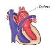
- A defect in which of the following aortic arches causes the defect shown in the image?
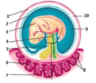
- Which of the following structures develops from the structure marked 4?
View More Questions
Top NEET PG Cervical plexus - greater auricular nerve Questions

Top NEET PG Questions
- A patient hailing from Delhi presents with fever, arthralgia, and extensive petechial rash for 3 days. Lab investigations revealed a hemoglobin of 9 g/ dL, a white blood cell count of 9000 cells/mm3, a platelet count of 20000 cells/mm3, and a prolonged bleeding time. The clotting time was normal. What is the most likely diagnosis?
- Which of the following statements is true about Trichomonas vaginalis?
- A lady from West Rajasthan presented with an ulcer surrounded by erythema on the right leg. Microscopy of the biopsy from the edge of the ulcer showed organisms with dark staining nuclei and kinetoplast. What is the most likely causative agent?
- A child presents with a fever and a rash. Urine examination showed cells with owl's eye appearance. What is the most likely diagnosis?
- Infection with Clonorchis sinensis is associated with an increased risk of__?
View More Questions