Question:
What is the stage of an ovarian cancer with bilateral involvement, capsular rupture, and positive ascitic fluid for malignant cells?
What is the stage of an ovarian cancer with bilateral involvement, capsular rupture, and positive ascitic fluid for malignant cells?
Show Hint
Capsule rupture plus positive ascitic cytology in a tumor confined to the ovaries defines FIGO stage IC, a substage of stage I.
Updated On: Jul 8, 2026
- I
- II
- III
- IV
Show Solution

Verified By Collegedunia
The Correct Option is A
Solution and Explanation
Step 1: Understanding the Question.
The case describes an ovarian cancer that involves both ovaries, has a ruptured capsule, and shows malignant cells in the ascitic fluid. We need to place this picture into the correct FIGO stage.
Step 2: Key Concept or Approach.
FIGO staging for ovarian cancer first asks whether the growth stays inside the ovaries or spreads out.
Stage I means the growth is limited to one or both ovaries. Within stage I:
Stage IA is one ovary, capsule intact, no tumor on the surface, no malignant cells in fluid.
Stage IB is both ovaries, capsule intact, no surface tumor, no malignant cells in fluid.
Stage IC is one or both ovaries with capsule rupture, tumor on the ovarian surface, or malignant cells found in the ascites or peritoneal washings.
Stage II adds spread within the pelvis, stage III adds spread beyond the pelvis or to lymph nodes, and stage IV means the cancer has reached distant organs.
Step 3: Detailed Explanation.
Here, both ovaries are involved but there is no mention of spread to the uterus, tubes, pelvic peritoneum, lymph nodes, or distant organs, so stages II, III, and IV do not fit this case.
The capsule rupture and the positive ascitic cytology are the two features that define stage IC, one of the three substages of stage I.
Bilateral disease alone does not push the stage higher, since the staging rests on where the tumor cells are found, not on how many ovaries carry the growth.
So this case sits inside stage I, specifically substage IC.
Step 4: Final Answer.
The disease is confined to the ovaries, with capsule rupture and positive ascitic cytology, which is FIGO stage I (substage IC).
\[ \boxed{\text{Stage I}} \]
The case describes an ovarian cancer that involves both ovaries, has a ruptured capsule, and shows malignant cells in the ascitic fluid. We need to place this picture into the correct FIGO stage.
Step 2: Key Concept or Approach.
FIGO staging for ovarian cancer first asks whether the growth stays inside the ovaries or spreads out.
Stage I means the growth is limited to one or both ovaries. Within stage I:
Stage IA is one ovary, capsule intact, no tumor on the surface, no malignant cells in fluid.
Stage IB is both ovaries, capsule intact, no surface tumor, no malignant cells in fluid.
Stage IC is one or both ovaries with capsule rupture, tumor on the ovarian surface, or malignant cells found in the ascites or peritoneal washings.
Stage II adds spread within the pelvis, stage III adds spread beyond the pelvis or to lymph nodes, and stage IV means the cancer has reached distant organs.
Step 3: Detailed Explanation.
Here, both ovaries are involved but there is no mention of spread to the uterus, tubes, pelvic peritoneum, lymph nodes, or distant organs, so stages II, III, and IV do not fit this case.
The capsule rupture and the positive ascitic cytology are the two features that define stage IC, one of the three substages of stage I.
Bilateral disease alone does not push the stage higher, since the staging rests on where the tumor cells are found, not on how many ovaries carry the growth.
So this case sits inside stage I, specifically substage IC.
Step 4: Final Answer.
The disease is confined to the ovaries, with capsule rupture and positive ascitic cytology, which is FIGO stage I (substage IC).
\[ \boxed{\text{Stage I}} \]
Was this answer helpful?
0
0
Top NEET PG Gynaecology and Obstetrics Questions
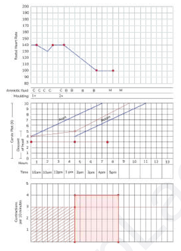
- A partogram of a woman who is in labor is shown below. Oxytocin infusion is already given. How will you manage the condition?
- A woman presents to you at 36 weeks of gestation with complaints of breathlessness and excessive abdominal distension. Fetal movements are normal. On examination, fetal parts are not easily felt and fetal heartbeat is heard but it is muffled. Her symphysis fundal height is 41 cm. Her abdomen is tense but not tender. What is the most likely diagonosis?
- For which of the following procedures in the given OT list, can you preferably do a hysteroscopy ?
- A primigravida presents to you with anemia early in her pregnancy. She is 7 weeks pregnant as seen on ultrasound. Her hemoglobin level is 9 g/dL. When should the iron supplements be started for her?
- A 23-year-old woman accompanied by her mother-in-law comes to the infertility clinic. She has been having regular intercourse for 6 months but is not able to conceive. What is the next best step?
View More Questions
Top NEET PG Questions
- A patient hailing from Delhi presents with fever, arthralgia, and extensive petechial rash for 3 days. Lab investigations revealed a hemoglobin of 9 g/ dL, a white blood cell count of 9000 cells/mm3, a platelet count of 20000 cells/mm3, and a prolonged bleeding time. The clotting time was normal. What is the most likely diagnosis?
- Which of the following statements is true about Trichomonas vaginalis?
- A lady from West Rajasthan presented with an ulcer surrounded by erythema on the right leg. Microscopy of the biopsy from the edge of the ulcer showed organisms with dark staining nuclei and kinetoplast. What is the most likely causative agent?
- A child presents with a fever and a rash. Urine examination showed cells with owl's eye appearance. What is the most likely diagnosis?
- Infection with Clonorchis sinensis is associated with an increased risk of__?
View More Questions