Question:
What is the diagnosis in a 65 year old patient with fever, flank pain, and calculi with fat densities?
What is the diagnosis in a 65 year old patient with fever, flank pain, and calculi with fat densities?
Show Hint
Fat-dense areas around an obstructing stone with chronic infection point to a specific destructive kidney disease.
Updated On: Jul 8, 2026
- Xanthogranulomatous pyelonephritis
- Renal abscess
- Chronic pyelonephritis
- Tuberculous kidney
Show Solution

Verified By Collegedunia
The Correct Option is A
Solution and Explanation
Step 1: Understanding the Question:
A 65 year old patient has fever, pain in the flank, and kidney stones that show fat density on imaging. We need to name the disease that fits this exact picture.
Step 2: Key Concept:
Xanthogranulomatous pyelonephritis (XGP) is a rare, severe form of chronic kidney infection. It destroys the kidney tissue and replaces it with lipid laden (fat filled) macrophages, called foam cells. This fat content is what shows up as fat density areas on CT scan. XGP is almost always linked to an obstructing stone, often a staghorn calculus, along with long standing infection, commonly by Proteus or E. coli.
Step 3: Detailed Explanation:
The three clues, fever, flank pain, and a stone with fat density, point straight to XGP. On CT, XGP classically shows an enlarged kidney with a central stone, dilated calyces filled with pus and debris, and surrounding fat dense material, sometimes called the "bear paw" sign.
Renal abscess also causes fever and flank pain, but it does not typically show a fat containing mass around a stone, it shows a fluid filled cavity with a thick wall.
Chronic pyelonephritis causes scarring and a small shrunken kidney with cortical thinning, not a fat density mass.
Tuberculous kidney causes calcification, cavitation, and a "putty kidney" in advanced cases, but not fat density change.
Step 4: Final Answer:
The fat density change around an obstructing calculus with fever and flank pain is the hallmark of xanthogranulomatous pyelonephritis.
A 65 year old patient has fever, pain in the flank, and kidney stones that show fat density on imaging. We need to name the disease that fits this exact picture.
Step 2: Key Concept:
Xanthogranulomatous pyelonephritis (XGP) is a rare, severe form of chronic kidney infection. It destroys the kidney tissue and replaces it with lipid laden (fat filled) macrophages, called foam cells. This fat content is what shows up as fat density areas on CT scan. XGP is almost always linked to an obstructing stone, often a staghorn calculus, along with long standing infection, commonly by Proteus or E. coli.
Step 3: Detailed Explanation:
The three clues, fever, flank pain, and a stone with fat density, point straight to XGP. On CT, XGP classically shows an enlarged kidney with a central stone, dilated calyces filled with pus and debris, and surrounding fat dense material, sometimes called the "bear paw" sign.
Renal abscess also causes fever and flank pain, but it does not typically show a fat containing mass around a stone, it shows a fluid filled cavity with a thick wall.
Chronic pyelonephritis causes scarring and a small shrunken kidney with cortical thinning, not a fat density mass.
Tuberculous kidney causes calcification, cavitation, and a "putty kidney" in advanced cases, but not fat density change.
Step 4: Final Answer:
The fat density change around an obstructing calculus with fever and flank pain is the hallmark of xanthogranulomatous pyelonephritis.
Was this answer helpful?
0
0
Top NEET PG Surgery Questions
- A 55-year-old male presented with verrucous carcinoma around the glans of the penis. Examination reveals that the inguinal lymph nodes are not enlarged. What is the appropriate management for this patient?
- Identify the procedure being performed in the image given below.
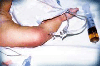
- A diabetic patient presents with sudden-onset perineal pain. On examination, foul-smelling discharge, and necrotic tissue as seen in the image is noted. Which of the following is true about the given condition?
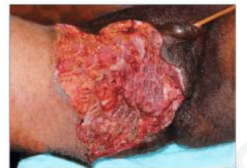
- Which of the following is the most common complication following ligation of the first vessel during abdominoperineal resection for rectal carcinoma?(APR Abdominoperineal resection)
- A patient presents to the clinic with the lesion given in the image. He had a traumatic injury to the chest one year ago. What is the most likely diagnosis?
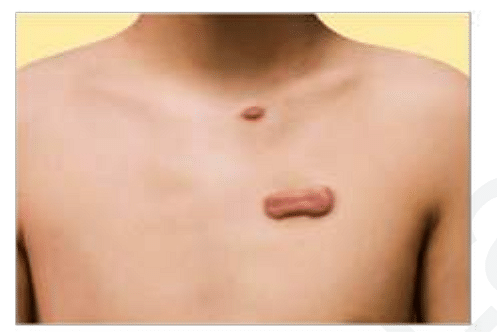
View More Questions
Top NEET PG Questions
- A patient hailing from Delhi presents with fever, arthralgia, and extensive petechial rash for 3 days. Lab investigations revealed a hemoglobin of 9 g/ dL, a white blood cell count of 9000 cells/mm3, a platelet count of 20000 cells/mm3, and a prolonged bleeding time. The clotting time was normal. What is the most likely diagnosis?
- Which of the following statements is true about Trichomonas vaginalis?
- A lady from West Rajasthan presented with an ulcer surrounded by erythema on the right leg. Microscopy of the biopsy from the edge of the ulcer showed organisms with dark staining nuclei and kinetoplast. What is the most likely causative agent?
- A child presents with a fever and a rash. Urine examination showed cells with owl's eye appearance. What is the most likely diagnosis?
- Infection with Clonorchis sinensis is associated with an increased risk of__?
View More Questions