Question:
Consider the following. Upper gastrointestinal endoscopy may reveal smooth mucosa in:
- Linitis plastica
- Lymphoma of stomach
- Stromal tumour
- Chronic atrophic gastritis
Which of these are correct?
Consider the following. Upper gastrointestinal endoscopy may reveal smooth mucosa in:
- Linitis plastica
- Lymphoma of stomach
- Stromal tumour
- Chronic atrophic gastritis
Which of these are correct?
Show Hint
Think about which lesions stay under the mucosa versus which ones tend to break through and ulcerate it.
Updated On: Jul 16, 2026
- 1 and 4
- 1 and 3
- 2 and 4
- 1, 2, 3 and 4
Show Solution

Verified By Collegedunia
The Correct Option is C
Solution and Explanation
Step 1: Understanding the Question:
The question lists four gastric conditions and asks which of them can show a smooth, rather than ulcerated or nodular, mucosal surface when viewed at upper GI endoscopy.
Step 2: Key Concept or Approach:
Endoscopy visualises only the surface mucosa. A lesion that is primarily submucosal or that diffusely thickens the wall without breaching the epithelium can leave the overlying mucosa looking smooth and largely intact, even though deeper biopsies show significant disease. A lesion that predominantly affects the mucosa itself, or that tends to ulcerate as it grows, is more likely to look irregular or eroded on endoscopy instead.
Step 3: Working Through the Options:
Gastric lymphoma frequently infiltrates the gastric wall submucosally and can spread widely while the mucosal surface over it stays smooth and only diffusely thickened, which is a recognised reason superficial endoscopic biopsies can under-sample and miss the diagnosis. Chronic atrophic gastritis flattens and effaces the normal rugal folds as the glands atrophy, so the mucosa looks thin, pale, and smooth rather than showing the usual rugose pattern, with submucosal vessels sometimes visible through it. Linitis plastica does infiltrate the stomach wall diffusely and can rigidify it, but it commonly shows patchy mucosal change such as erythema, ulceration, or nodularity rather than a uniformly smooth surface, which is exactly why endoscopic biopsy yield in linitis plastica is notoriously poor. A gastrointestinal stromal tumour usually presents as a discrete submucosal bulge, and once it outgrows its blood supply it very often ulcerates centrally, giving an umbilicated rather than a uniformly smooth appearance. Taking these features together, lymphoma of the stomach and chronic atrophic gastritis are the two conditions most consistently associated with a smooth endoscopic mucosal appearance.
Step 4: Conclusion:
The correct combination is gastric lymphoma and chronic atrophic gastritis.
The question lists four gastric conditions and asks which of them can show a smooth, rather than ulcerated or nodular, mucosal surface when viewed at upper GI endoscopy.
Step 2: Key Concept or Approach:
Endoscopy visualises only the surface mucosa. A lesion that is primarily submucosal or that diffusely thickens the wall without breaching the epithelium can leave the overlying mucosa looking smooth and largely intact, even though deeper biopsies show significant disease. A lesion that predominantly affects the mucosa itself, or that tends to ulcerate as it grows, is more likely to look irregular or eroded on endoscopy instead.
Step 3: Working Through the Options:
Gastric lymphoma frequently infiltrates the gastric wall submucosally and can spread widely while the mucosal surface over it stays smooth and only diffusely thickened, which is a recognised reason superficial endoscopic biopsies can under-sample and miss the diagnosis. Chronic atrophic gastritis flattens and effaces the normal rugal folds as the glands atrophy, so the mucosa looks thin, pale, and smooth rather than showing the usual rugose pattern, with submucosal vessels sometimes visible through it. Linitis plastica does infiltrate the stomach wall diffusely and can rigidify it, but it commonly shows patchy mucosal change such as erythema, ulceration, or nodularity rather than a uniformly smooth surface, which is exactly why endoscopic biopsy yield in linitis plastica is notoriously poor. A gastrointestinal stromal tumour usually presents as a discrete submucosal bulge, and once it outgrows its blood supply it very often ulcerates centrally, giving an umbilicated rather than a uniformly smooth appearance. Taking these features together, lymphoma of the stomach and chronic atrophic gastritis are the two conditions most consistently associated with a smooth endoscopic mucosal appearance.
Step 4: Conclusion:
The correct combination is gastric lymphoma and chronic atrophic gastritis.
Was this answer helpful?
0
0
Top NEET PG Surgery Questions
- A 55-year-old male presented with verrucous carcinoma around the glans of the penis. Examination reveals that the inguinal lymph nodes are not enlarged. What is the appropriate management for this patient?
- Identify the procedure being performed in the image given below.

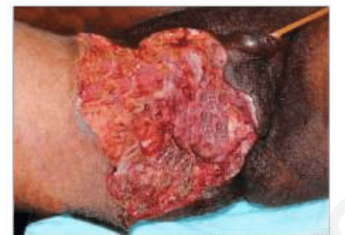
- A diabetic patient presents with sudden-onset perineal pain. On examination, foul-smelling discharge, and necrotic tissue as seen in the image is noted. Which of the following is true about the given condition?
- Which of the following is the most common complication following ligation of the first vessel during abdominoperineal resection for rectal carcinoma?(APR Abdominoperineal resection)
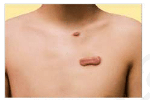
- A patient presents to the clinic with the lesion given in the image. He had a traumatic injury to the chest one year ago. What is the most likely diagnosis?
View More Questions
Top NEET PG Questions
- A patient hailing from Delhi presents with fever, arthralgia, and extensive petechial rash for 3 days. Lab investigations revealed a hemoglobin of 9 g/ dL, a white blood cell count of 9000 cells/mm3, a platelet count of 20000 cells/mm3, and a prolonged bleeding time. The clotting time was normal. What is the most likely diagnosis?
- Which of the following statements is true about Trichomonas vaginalis?
- A lady from West Rajasthan presented with an ulcer surrounded by erythema on the right leg. Microscopy of the biopsy from the edge of the ulcer showed organisms with dark staining nuclei and kinetoplast. What is the most likely causative agent?
- A child presents with a fever and a rash. Urine examination showed cells with owl's eye appearance. What is the most likely diagnosis?
- Infection with Clonorchis sinensis is associated with an increased risk of__?
View More Questions