Question:
A patient has Proteus infection and now plain xray of abdomen was done and a large stone was shown in urinary bladder. Stone is made up of
A patient has Proteus infection and now plain xray of abdomen was done and a large stone was shown in urinary bladder. Stone is made up of
Show Hint
Proteus = urease = alkaline urine = phosphate (infection) stone. Pick the phosphate option.
Updated On: Aug 27, 2026
- Calcium Phosphate
- Cysteine
- Calcium Oxalate
- Xanthine
Show Solution

Verified By Collegedunia
The Correct Option is A
Solution and Explanation
Step 1: Identify the key clue. The patient has a Proteus urinary infection. Proteus mirabilis is a urease-producing organism - this single fact dictates the stone type.
Step 2: The urease mechanism. Urease splits urinary urea into ammonia and carbon dioxide: \[\text{Urea} + \text{H}_2\text{O} \xrightarrow{\text{urease}} 2\,\text{NH}_3 + \text{CO}_2\] The ammonia raises (alkalinises) the urine pH. In persistently alkaline urine, magnesium, ammonium and phosphate precipitate.
Step 3: The stone formed. Urease-producing organisms classically form struvite stones - magnesium ammonium phosphate, often mixed with calcium phosphate (carbonate-apatite). These are the "infection (triple-phosphate) stones" and tend to grow large, filling the collecting system (staghorn) or, as here, forming a large bladder stone. They are radio-opaque, consistent with being seen on plain X-ray.
Step 4: Why option A (Calcium Phosphate) is correct. Among the four options, only calcium phosphate is a phosphate, alkaline-urine, infection-related stone consistent with a urease-positive Proteus infection. It is the answer key choice for the phosphate/infection stone.
Step 5: Why the distractors are wrong. (B) Cystine (cysteine) stones occur in cystinuria, a hereditary tubular transport defect, unrelated to infection; urine tends to be acidic. (C) Calcium oxalate stones form in normal-to-acidic urine from metabolic/dietary causes, not from urease-driven alkalinisation. (D) Xanthine stones are rare, due to xanthine oxidase deficiency or allopurinol therapy, and are radiolucent - not infection-related.
Final Answer: Option A - Calcium Phosphate.
Step 2: The urease mechanism. Urease splits urinary urea into ammonia and carbon dioxide: \[\text{Urea} + \text{H}_2\text{O} \xrightarrow{\text{urease}} 2\,\text{NH}_3 + \text{CO}_2\] The ammonia raises (alkalinises) the urine pH. In persistently alkaline urine, magnesium, ammonium and phosphate precipitate.
Step 3: The stone formed. Urease-producing organisms classically form struvite stones - magnesium ammonium phosphate, often mixed with calcium phosphate (carbonate-apatite). These are the "infection (triple-phosphate) stones" and tend to grow large, filling the collecting system (staghorn) or, as here, forming a large bladder stone. They are radio-opaque, consistent with being seen on plain X-ray.
Step 4: Why option A (Calcium Phosphate) is correct. Among the four options, only calcium phosphate is a phosphate, alkaline-urine, infection-related stone consistent with a urease-positive Proteus infection. It is the answer key choice for the phosphate/infection stone.
Step 5: Why the distractors are wrong. (B) Cystine (cysteine) stones occur in cystinuria, a hereditary tubular transport defect, unrelated to infection; urine tends to be acidic. (C) Calcium oxalate stones form in normal-to-acidic urine from metabolic/dietary causes, not from urease-driven alkalinisation. (D) Xanthine stones are rare, due to xanthine oxidase deficiency or allopurinol therapy, and are radiolucent - not infection-related.
Final Answer: Option A - Calcium Phosphate.
Was this answer helpful?
0
0
Top NEET PG Surgery Questions
- A 55-year-old male presented with verrucous carcinoma around the glans of the penis. Examination reveals that the inguinal lymph nodes are not enlarged. What is the appropriate management for this patient?
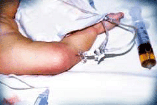
- Identify the procedure being performed in the image given below.
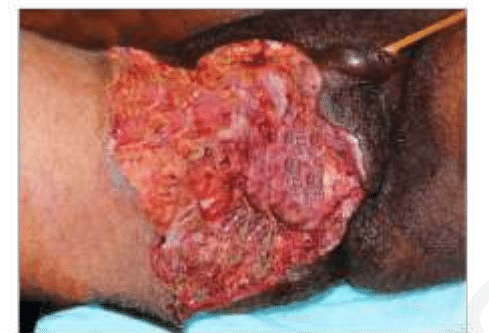
- A diabetic patient presents with sudden-onset perineal pain. On examination, foul-smelling discharge, and necrotic tissue as seen in the image is noted. Which of the following is true about the given condition?
- Which of the following is the most common complication following ligation of the first vessel during abdominoperineal resection for rectal carcinoma?(APR Abdominoperineal resection)
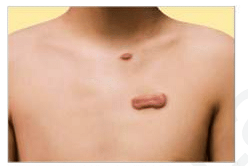
- A patient presents to the clinic with the lesion given in the image. He had a traumatic injury to the chest one year ago. What is the most likely diagnosis?
View More Questions
Top NEET PG Questions
- A patient hailing from Delhi presents with fever, arthralgia, and extensive petechial rash for 3 days. Lab investigations revealed a hemoglobin of 9 g/ dL, a white blood cell count of 9000 cells/mm3, a platelet count of 20000 cells/mm3, and a prolonged bleeding time. The clotting time was normal. What is the most likely diagnosis?
- Which of the following statements is true about Trichomonas vaginalis?
- A lady from West Rajasthan presented with an ulcer surrounded by erythema on the right leg. Microscopy of the biopsy from the edge of the ulcer showed organisms with dark staining nuclei and kinetoplast. What is the most likely causative agent?
- A child presents with a fever and a rash. Urine examination showed cells with owl's eye appearance. What is the most likely diagnosis?
- Infection with Clonorchis sinensis is associated with an increased risk of__?
View More Questions