Question:
A 16 year old girl failed her final examination. Disgusted with life, she cut across the front of her wrist at the flexor retinaculum. She was rushed to hospital, and the surgeon noticed that the cut was superficial. All of the following structures would have been damaged except:
A 16 year old girl failed her final examination. Disgusted with life, she cut across the front of her wrist at the flexor retinaculum. She was rushed to hospital, and the surgeon noticed that the cut was superficial. All of the following structures would have been damaged except:
Show Hint
Ask which of these structures sits deep to the flexor retinaculum inside the carpal tunnel, that one is protected from a superficial cut.
Updated On: Jul 8, 2026
- Ulnar nerve
- Median nerve
- Palmar cutaneous branch of median nerve
- Superficial branch of radial artery
Show Solution

Verified By Collegedunia
The Correct Option is B
Solution and Explanation
Step 1: Picture the layers at the front of the wrist.
At the wrist, the flexor retinaculum is a tough band that roofs over the carpal tunnel. Some structures lie deep to it, inside the carpal tunnel, and some lie superficial to it, just under the skin. A superficial cut only reaches the structures in that shallow, superficial layer.
Step 2: Place the ulnar nerve.
The ulnar nerve does not pass through the carpal tunnel. It runs superficial to the flexor retinaculum, through Guyon's canal, alongside the ulnar artery, on the medial side of the wrist. A superficial cut across the wrist would injure it.
Step 3: Place the palmar cutaneous branch of the median nerve.
This branch leaves the main median nerve a little above the wrist and travels superficial to the flexor retinaculum to supply skin over the base of the palm. It is superficial, so a shallow cut would injure it too.
Step 4: Place the superficial branch of the radial artery.
The superficial palmar branch of the radial artery crosses the wrist superficially, often over or through the thenar muscles, to help complete the superficial palmar arch. It lies in the superficial plane, so a superficial cut would damage it as well.
Step 5: Place the median nerve trunk itself.
Unlike its cutaneous branch, the main trunk of the median nerve travels deep to the flexor retinaculum, inside the carpal tunnel, together with the nine flexor tendons. Because the surgeon specifically noted the cut was superficial, it would not have reached the median nerve trunk lying deep to the retinaculum.
Step 6: Final conclusion.
The structure that would NOT be damaged by a superficial wrist cut is the median nerve itself.
\[ \boxed{\text{Median nerve}} \]
At the wrist, the flexor retinaculum is a tough band that roofs over the carpal tunnel. Some structures lie deep to it, inside the carpal tunnel, and some lie superficial to it, just under the skin. A superficial cut only reaches the structures in that shallow, superficial layer.
Step 2: Place the ulnar nerve.
The ulnar nerve does not pass through the carpal tunnel. It runs superficial to the flexor retinaculum, through Guyon's canal, alongside the ulnar artery, on the medial side of the wrist. A superficial cut across the wrist would injure it.
Step 3: Place the palmar cutaneous branch of the median nerve.
This branch leaves the main median nerve a little above the wrist and travels superficial to the flexor retinaculum to supply skin over the base of the palm. It is superficial, so a shallow cut would injure it too.
Step 4: Place the superficial branch of the radial artery.
The superficial palmar branch of the radial artery crosses the wrist superficially, often over or through the thenar muscles, to help complete the superficial palmar arch. It lies in the superficial plane, so a superficial cut would damage it as well.
Step 5: Place the median nerve trunk itself.
Unlike its cutaneous branch, the main trunk of the median nerve travels deep to the flexor retinaculum, inside the carpal tunnel, together with the nine flexor tendons. Because the surgeon specifically noted the cut was superficial, it would not have reached the median nerve trunk lying deep to the retinaculum.
Step 6: Final conclusion.
The structure that would NOT be damaged by a superficial wrist cut is the median nerve itself.
\[ \boxed{\text{Median nerve}} \]
Was this answer helpful?
0
0
Top NEET PG Anatomy Questions
- A defect in which of the following forms the structure marked below?

- A student had his jaw locked while yawning. Which of the following muscles is attached to the articular disc of the temporomandibular joint?
- The given histology image is of ?

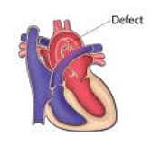
- A defect in which of the following aortic arches causes the defect shown in the image?
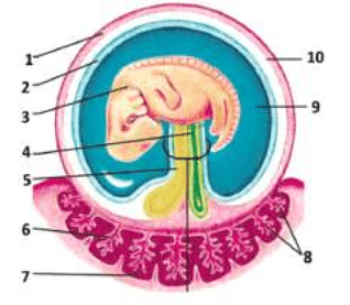
- Which of the following structures develops from the structure marked 4?
View More Questions
Top NEET PG Questions
- A patient hailing from Delhi presents with fever, arthralgia, and extensive petechial rash for 3 days. Lab investigations revealed a hemoglobin of 9 g/ dL, a white blood cell count of 9000 cells/mm3, a platelet count of 20000 cells/mm3, and a prolonged bleeding time. The clotting time was normal. What is the most likely diagnosis?
- Which of the following statements is true about Trichomonas vaginalis?
- A lady from West Rajasthan presented with an ulcer surrounded by erythema on the right leg. Microscopy of the biopsy from the edge of the ulcer showed organisms with dark staining nuclei and kinetoplast. What is the most likely causative agent?
- A child presents with a fever and a rash. Urine examination showed cells with owl's eye appearance. What is the most likely diagnosis?
- Infection with Clonorchis sinensis is associated with an increased risk of__?
View More Questions