Question:
A 12-year-old child is admitted to the ICU with blunt trauma and a femur fracture. PaO2 stays at 60 mmHg despite 100% oxygen via a non-rebreather mask, the chest X-ray shows clear lung fields, but the patient remains confused. What is the most likely diagnosis?
A 12-year-old child is admitted to the ICU with blunt trauma and a femur fracture. PaO2 stays at 60 mmHg despite 100% oxygen via a non-rebreather mask, the chest X-ray shows clear lung fields, but the patient remains confused. What is the most likely diagnosis?
Show Hint
Long-bone fracture, hypoxia and confusion with a clear CXR points where?
Updated On: Jun 23, 2026
- Pulmonary contusion
- Fat embolism syndrome
- Hypovolaemic shock
- Pulmonary embolism
Show Solution

Verified By Collegedunia
The Correct Option is B
Solution and Explanation
Answer: Fat embolism syndrome.
Step 1: The setting is a long-bone (femur) fracture with two organ systems hit: the lungs (hypoxia, PaO2 60 mmHg despite 100% oxygen) and the brain (confusion). This combination after a long-bone fracture points to fat embolism syndrome (FES).
Step 2: Fat globules released from the marrow lodge in capillary beds, causing direct tissue injury and a systemic inflammatory response. This produces the classic triad: respiratory failure, neurological changes and a petechial rash. Onset is typically 24 to 72 hours after the fracture.
Step 3: A clear chest X-ray with severe refractory hypoxia argues against pulmonary contusion, which would show lung opacities, and against the volume-loss picture of hypovolaemic shock. Pulmonary embolism (thrombotic) is far less likely in a child right after a fracture and does not explain the marrow-fat origin and CNS picture as well.
Step 4: Hypoxia plus confusion after a femur fracture equals fat embolism syndrome. Ref: Bailey and Love / Schwartz Principles of Surgery.
Step 1: The setting is a long-bone (femur) fracture with two organ systems hit: the lungs (hypoxia, PaO2 60 mmHg despite 100% oxygen) and the brain (confusion). This combination after a long-bone fracture points to fat embolism syndrome (FES).
Step 2: Fat globules released from the marrow lodge in capillary beds, causing direct tissue injury and a systemic inflammatory response. This produces the classic triad: respiratory failure, neurological changes and a petechial rash. Onset is typically 24 to 72 hours after the fracture.
Step 3: A clear chest X-ray with severe refractory hypoxia argues against pulmonary contusion, which would show lung opacities, and against the volume-loss picture of hypovolaemic shock. Pulmonary embolism (thrombotic) is far less likely in a child right after a fracture and does not explain the marrow-fat origin and CNS picture as well.
Step 4: Hypoxia plus confusion after a femur fracture equals fat embolism syndrome. Ref: Bailey and Love / Schwartz Principles of Surgery.
Was this answer helpful?
0
0
Top NEET PG Surgery Questions
- A 55-year-old male presented with verrucous carcinoma around the glans of the penis. Examination reveals that the inguinal lymph nodes are not enlarged. What is the appropriate management for this patient?
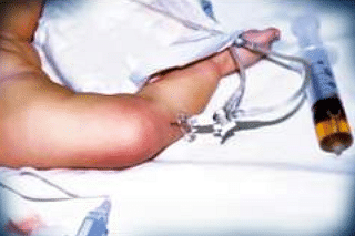
- Identify the procedure being performed in the image given below.
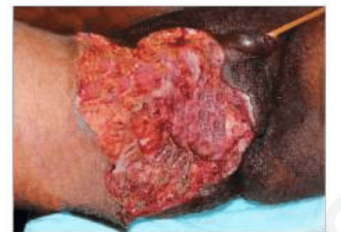
- A diabetic patient presents with sudden-onset perineal pain. On examination, foul-smelling discharge, and necrotic tissue as seen in the image is noted. Which of the following is true about the given condition?
- Which of the following is the most common complication following ligation of the first vessel during abdominoperineal resection for rectal carcinoma?(APR Abdominoperineal resection)
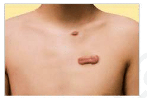
- A patient presents to the clinic with the lesion given in the image. He had a traumatic injury to the chest one year ago. What is the most likely diagnosis?
View More Questions
Top NEET PG Fat embolism syndrome Questions
Top NEET PG Questions
- A patient hailing from Delhi presents with fever, arthralgia, and extensive petechial rash for 3 days. Lab investigations revealed a hemoglobin of 9 g/ dL, a white blood cell count of 9000 cells/mm3, a platelet count of 20000 cells/mm3, and a prolonged bleeding time. The clotting time was normal. What is the most likely diagnosis?
- Which of the following statements is true about Trichomonas vaginalis?
- A lady from West Rajasthan presented with an ulcer surrounded by erythema on the right leg. Microscopy of the biopsy from the edge of the ulcer showed organisms with dark staining nuclei and kinetoplast. What is the most likely causative agent?
- A child presents with a fever and a rash. Urine examination showed cells with owl's eye appearance. What is the most likely diagnosis?
- Infection with Clonorchis sinensis is associated with an increased risk of__?
View More Questions